
What Is the Maxilla?
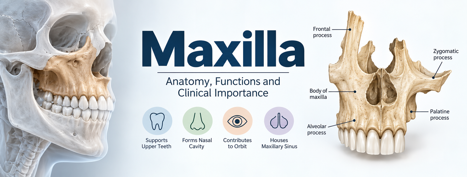
The maxilla is the bone that forms the upper jaw and is one of the most important bones of the human facial skeleton. It supports the upper teeth and contributes to the structure of the mouth, nasal cavity, hard palate, and eye socket (orbit).
The maxilla is actually a paired bone. There is a right maxilla and a left maxilla, and the two meet at the midline. Together, they form the upper jaw. Unlike the mandible, which is the movable lower jaw, the maxilla is part of the fixed facial skeleton.
The maxilla also contains the maxillary sinus, one of the major paranasal sinuses, making this bone important not only in dentistry and facial anatomy but also in sinus and maxillofacial conditions.
Quick facts about the maxilla
| Feature | Description |
|---|---|
| Common name | Upper jaw bone |
| Number | Two bones — right and left |
| Location | Central part of the facial skeleton |
| Main role | Supports upper teeth and forms part of the midface |
| Major cavity | Maxillary sinus |
| Processes | Frontal, zygomatic, alveolar and palatine |
| Major structures formed | Orbit, nasal cavity and hard palate |
| Related field | Dentistry, anatomy, oral surgery and maxillofacial surgery |
Maxilla Anatomy
Understanding maxilla anatomy becomes easier when the bone is divided into its main structural components.
The adult maxilla consists of a body and four processes:
- Frontal process
- Zygomatic process
- Alveolar process
- Palatine process
The body of the maxilla is roughly pyramidal and contains the maxillary sinus.

1. Body of the Maxilla
The body of the maxilla is the central portion of the bone. It contains the maxillary sinus, a hollow air-filled space within the facial skeleton.
The body has surfaces that relate to several important regions of the face, including the orbit, nasal cavity and infratemporal region.
Because of its position, the body of the maxilla is involved in several important anatomical relationships and clinical procedures.
2. Frontal Process of the Maxilla
The frontal process projects upward from the maxilla toward the frontal region of the skull.
It contributes to the side of the nasal region and forms part of the boundary between the nasal and orbital areas. The process also articulates with neighboring facial bones.
The frontal process is therefore important in understanding the bony framework of the nose, orbit and midface.
3. Zygomatic Process of the Maxilla
The zygomatic process extends laterally from the maxilla and connects with the zygomatic bone, commonly associated with the cheekbone.
This region helps provide structural support to the midface and participates in the transmission of forces generated during chewing.
4. Alveolar Process of the Maxilla
The alveolar process forms the part of the maxilla that surrounds and supports the roots of the upper teeth.
It contains the dental alveoli, or tooth sockets, where the upper teeth are anchored.
This makes the alveolar process particularly important in:
- Dentistry
- Tooth extraction
- Dental implants
- Orthodontics
- Periodontal treatment
- Oral surgery
The maxillary alveolar region also changes over time in response to tooth development, eruption and tooth loss.
5. Palatine Process of the Maxilla
The palatine process extends medially from the maxilla and contributes substantially to the hard palate, which forms the bony roof of the mouth.
The hard palate separates the oral cavity from the nasal cavity and plays an important role in eating, swallowing and speech.
The two maxillae meet at the midline, contributing to the bony structure of the anterior palate.
Important Anatomical Landmarks of the Maxilla
Several openings, grooves, projections and surfaces make the maxilla a complex bone.
Important maxillary landmarks include:
- Infraorbital foramen
- Infraorbital canal
- Infraorbital groove
- Maxillary sinus
- Anterior nasal spine
- Nasal crest
- Nasal notch
- Canine fossa
- Maxillary tuberosity
- Incisive canal
- Incisive foramen
- Alveolar process
- Frontal process
- Zygomatic process
- Palatine process
Anatomy Standard similarly organizes the maxilla around its surfaces, processes, sinus, foramina and other bony landmarks.
Infraorbital Foramen
The infraorbital foramen is an opening located below the orbit.
It provides a passage for the infraorbital nerve and accompanying blood vessels. Because of its anatomical location, it is an important landmark in dental anesthesia, facial surgery and procedures involving the midface.
Maxillary Sinus
The maxillary sinus is an air-filled cavity located inside the body of the maxilla.
It is one of the paranasal sinuses and is clinically important because of its close relationship with the upper teeth and surrounding structures.
The relationship between the maxillary sinus and upper posterior teeth is especially important in dentistry. Dental procedures involving upper molars and premolars may require careful consideration of the sinus because tooth roots can be close to its floor.
Anterior Nasal Spine
The anterior nasal spine is a bony projection in the anterior midline of the maxilla.
It contributes to the structural framework of the nose and is an important landmark when studying the relationship between the maxilla and nasal cavity.
Maxillary Tuberosity
The maxillary tuberosity is the rounded posterior portion of the maxilla behind the last upper molar region.
It is an important anatomical landmark in dental and oral surgical procedures.
What Does the Maxilla Do?
The maxilla has several important functions. It is not simply a bone that holds the upper teeth.
Major functions of the maxilla include:
1. Supports the upper teeth
The alveolar process contains the sockets that hold the upper teeth.
2. Forms part of the face
The maxilla contributes substantially to the structure and appearance of the middle third of the face.
3. Forms part of the orbit
The maxilla contributes to the bony structure of the eye socket.
4. Contributes to the nasal cavity
Parts of the maxilla form boundaries of the nasal region.
5. Forms part of the hard palate
Its palatine processes contribute to the roof of the mouth.
6. Houses the maxillary sinus
The body of the maxilla contains the maxillary sinus.
7. Helps transmit chewing forces
The maxilla participates in the structural framework that transfers forces generated during mastication to the facial skeleton.
Maxilla and Teeth
One of the most important functions of the maxilla is its relationship with the upper teeth.
The alveolar process contains tooth sockets for the maxillary teeth. These sockets provide the bony foundation necessary to anchor the teeth.
In adults, the upper dental arch normally contains:
- 4 incisors
- 2 canines
- 4 premolars
- 6 molars
for a total of 16 upper teeth when all permanent teeth, including third molars, are present.
The condition of the maxillary bone and its surrounding tissues is therefore highly relevant to dental health.
Maxilla vs Mandible
A common anatomy question is the difference between the maxilla and mandible.
| Maxilla | Mandible |
|---|---|
| Forms the upper jaw | Forms the lower jaw |
| Paired bone | Single fused bone in the adult |
| Generally fixed | Movable |
| Supports upper teeth | Supports lower teeth |
| Contains maxillary sinuses | Does not contain a comparable paranasal sinus |
| Forms part of orbit | Does not form the orbit |
| Contributes to hard palate | Does not form the hard palate |
The mandible is the movable jaw, while the maxilla forms the fixed upper jaw and contributes to several other structures of the facial skeleton.
Bones That Articulate With the Maxilla
The maxilla is highly connected to surrounding bones.
Each maxilla articulates with numerous neighboring bones, including the:
- Frontal bone
- Ethmoid bone
- Nasal bone
- Zygomatic bone
- Lacrimal bone
- Palatine bone
- Vomer
- Inferior nasal concha
- Opposite maxilla
This extensive articulation helps explain why the maxilla is considered an important structural component of the midface.
Blood Supply and Nerve Relationships
The maxilla is closely associated with important nerves and blood vessels.
Clinically significant structures include branches associated with the maxillary artery and sensory branches of the maxillary division of the trigeminal nerve (V2).
Important nerves associated with the maxilla include branches supplying:
- Upper teeth
- Gingiva
- Midface
- Nasal structures
- Palate
The infraorbital nerve is particularly important because it passes through the infraorbital canal and exits through the infraorbital foramen.
These relationships are important for local anesthesia, dental surgery and maxillofacial procedures.
Development of the Maxilla
The maxilla develops during embryonic growth through intramembranous ossification.
Development begins during early prenatal development, with ossification centers appearing around the sixth week. The developing structures subsequently unite as facial development progresses.
The maxillary sinus also develops during early life and continues to enlarge as the facial skeleton develops. Its size and shape change considerably during childhood and later development.
Normal development of the maxilla is essential for:
- Facial proportions
- Nasal development
- Dental arch development
- Palate formation
- Proper relationship between the upper and lower jaws
Abnormal development can contribute to craniofacial conditions that may require orthodontic, dental or surgical management.
Clinical Importance of the Maxilla
Because the maxilla is connected to the teeth, nasal cavity, orbit and maxillary sinus, disorders or injuries involving this bone can have consequences beyond the jaw itself.
Maxillary Fractures
Trauma to the face can result in fractures of the maxilla.
One important classification system is the Le Fort classification, which describes characteristic patterns of midface fractures.
The three classic patterns are:
- Le Fort I
- Le Fort II
- Le Fort III
These fractures can involve different portions of the maxilla and surrounding facial structures.
Maxillary injuries can potentially affect chewing, speech, breathing, facial structure and dental alignment, depending on their location and severity.
Dental Procedures and the Maxilla
The anatomy of the maxilla is especially important during:
- Tooth extraction
- Dental implant placement
- Root canal treatment
- Orthodontic treatment
- Bone grafting
- Sinus-related dental procedures
- Local anesthesia
- Oral and maxillofacial surgery
For example, the relationship between the maxillary sinus and posterior upper teeth must be considered during certain dental extractions and implant procedures.
Common Conditions Associated With the Maxilla
A variety of conditions can involve the maxilla or structures immediately surrounding it.
These may include:
Maxillary fractures
Trauma can fracture the maxilla and disrupt the normal structure of the midface.
Maxillary sinusitis
Because the maxillary sinus is located inside the maxilla, inflammation of this sinus can produce pain or pressure in the cheek and upper tooth region.
Dental infections
Infections involving upper teeth can sometimes spread toward adjacent structures, including the maxillary sinus.
Developmental abnormalities
Altered development of the maxilla can influence facial proportions, occlusion and dental alignment.
Maxillary tumors and cysts
Pathological lesions can arise in or around the maxilla and may affect adjacent structures.
Because the maxilla is anatomically connected to several important regions, diagnosis often requires a combination of clinical examination and appropriate imaging.
Why Is the Maxilla Important in Dentistry?
For dental professionals, the maxilla is one of the most clinically important bones of the skull.
Its anatomy affects:
- Tooth positioning
- Dental occlusion
- Local anesthesia
- Implant placement
- Tooth extraction
- Periodontal procedures
- Orthodontic movement
- Sinus-related complications
- Oral surgery
The maxilla’s foramina and canals also provide pathways for nerves and vessels that must be considered when planning dental procedures.
Maxilla in Orthodontics and Facial Development
The position and growth of the maxilla influence the relationship between the upper and lower teeth.
If the maxilla is positioned too far forward, too far backward, too narrow or otherwise develops differently from expected, it can contribute to malocclusion.
Orthodontists may evaluate:
- Maxillary width
- Maxillary position
- Upper dental arch
- Relationship between maxilla and mandible
- Facial growth pattern
- Tooth alignment
In selected cases, orthodontic treatment or orthognathic surgery may be used to improve the relationship between the jaws.
Maxilla in Oral and Maxillofacial Surgery
The complex anatomy of the maxilla makes it particularly important in oral and maxillofacial surgery.
Surgical planning may need to account for the:
- Orbit
- Nasal cavity
- Maxillary sinus
- Infraorbital nerve
- Upper teeth
- Palate
- Major facial blood vessels
- Adjacent facial bones
The maxilla is also involved in reconstructive procedures and orthognathic operations designed to correct jaw position and facial relationships.
Frequently Asked Questions About the Maxilla
Is the maxilla the upper jaw?
Yes. The maxilla is the bone that forms the upper jaw. It supports the upper teeth and also contributes to the orbit, nasal cavity and hard palate.
Is the maxilla one bone or two?
The maxilla develops as a paired structure, consisting of right and left maxillary bones that meet at the midline.
What are the four processes of the maxilla?
The four major processes are:
- Frontal process
- Zygomatic process
- Alveolar process
- Palatine process
What is the main function of the maxilla?
The maxilla supports the upper teeth and contributes to the structure of the midface, orbit, nasal cavity and hard palate. It also contains the maxillary sinus.
Does the maxilla move?
Unlike the mandible, the maxilla is part of the fixed facial skeleton and does not normally move during chewing.
Where is the maxillary sinus located?
The maxillary sinus is located inside the body of the maxilla, beneath the orbit and adjacent to the nasal cavity.
What is the difference between maxilla and mandible?
The maxilla forms the upper jaw, while the mandible forms the lower jaw. The mandible is movable, whereas the maxilla is fixed as part of the facial skeleton.
Why is the maxilla important for dentists?
The maxilla contains the sockets for the upper teeth and is closely related to important nerves, blood vessels, the nasal cavity and maxillary sinus. This makes its anatomy essential for dental treatment and oral surgery.